Okay, I can't take the credit for the clever title, or the suggestion for the topic... they were given to me courtesy of one of my students (soon to be graduates), Matt. Kudos to Matt!
This post is dedicated to practical tidbits of information that contribute to the performance of a smooth BE. I know when I graduated, I didn't feel that comfortable performing them, and then all of a sudden I was expected to be a barium-slinging super-tech once I got the "RT(R)" behind my name. I hope some if this helps.
A thorough history is important for BE's. I like to double check to see if the patient has had an endoscopy recently, or if they have had a recent biopsy. Depending on your hospital's routine for waiting after a biopsy, you may not do the procedure, or it will change to single contrast due to risk of perforation.
Ask about usage of laxatives... if there has been long-term use, the patient may very well have increased elasticity of the large intestine to a condition called megacolon. It would be a good idea to have some additional barium and/or supplies ready if this is the case.
Another thing to ask the patient before the exam is whether or not they followed their prep. Check with your hospital to see if they require an enema, laxative, or a combination of both (with NPO instructions of course). The stool should be clear without any particles in it.
When mixing your barium bag, make sure you use luke-warm water for single contrast. If you are using pre-mixed barium for a double, then you can place it on top of a processor for about 20 minutes before the patient arrives to warm it. Or you could place the bag in the sink and let warm water run all around it. Which ever way you do it, the warm water is supposed to be less of a shock to the colon upon entry. The colon will spasm less during your radiographs, and it can contribute to patient comfort. Hey, every little thing helps, right?
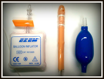
Connect the balloon inflator, and always test the balloon by inflating it outside the patient. It is rare, but there can be manufacturer defects on basically any type of mass-produced supply, and you don't want the balloon failing on you in the middle of the procedure.
I always like to prepare to do a double-inflation. You can do this on one single squeeze of the inflator. Just keep the tube clamped during your test inflation while pulling the inflator off of the tube. Let the inflator re-gain its original shape, then re-attach it to the tube. Take the clamp off the tube, and when the balloon deflates, you will now have approximately "two puffs" or twice the amount of air required to inflate the balloon available in one squeeze. The inflator will be quite distended. This should be done with caution... never give more than one inflation of air without a Radiologist's approval. This method should only be done while inflating the balloon under fluoroscopic guidance (which is routine at my current facility). The Radiologist usually tells us when to stop inflating during the procedure. RISK OF PERFORATION OF THE COLON increases if not done under fluoro with Radiologist supervision.
I also like to place one piece of tape around all of the tubes in order to make the bag assembly easier to handle while the patient is turning during the procedure. have a strip of tape torn off and placed on the x-ray table so that after the balloon is inflated, you can apply this tape about 2 inches away from the inflator, and bind the inflator tube, the barium tube, and the blue tube (for giving the patient air) all together.
Another thing I've noticed is how poorly the clamps work on the barium tubing. Occasionally, the Radiologist will be inserting air into the bowel (by squeezing the blue bulb), and you will not be able to see air entering the intestine on the fluoro screen for quite some time. What is happening is that the air is escaping around the clamp on the tube and going into the bag. I will bet you a dollar that if you turn around and look at your barium bag, you'll be able to see it inflating while everyone else is watching the fluoro monitor! The bowel will receive the air eventually, but only after the bag has filled with air, and the pressure in the bag is greater than the pressure required for it to go into the colon. Carry some hemostats with you. I like to bend the tube, or put a kink in it, and place a hemostat over the tube in addition to the existing clamp. This will prevent air leakage into the bag.
During the Radiologist's fluoro routine, you should be manning the barium flow from the bag into the patient. I would suggest instead of using the clamp to leave the clamp open and pinch the tube with your fingers to control the flow. This allows you to stop the flow quickly during exposures by the Radiologist. You should attempt to stop the flow momentarily during the exposure to prevent motion on the film. Also, the barium should not flow in too fast, or you will risk perforation, and you should never let it flow beyond your field of view on the fluoro screen. You may never know you caused a perforation until the tower is moved, or the barium extravasates into your field of view. Sometimes, even if it does not perforate, it can go far into the small intestine before you even realize it, and that is not desirable for a good diagnostic study.
During the study, we radiographers wear many hats. We are responsible for the flow of the barium, assisting with patient movement/rotation, and changing films out of the fluoro tower (if you're still using a spot camera). We should also be keeping an eye on the table. If you are running barium into the patient and keep watching the fluoro monitor anxiously waiting to see it rush in, make sure it's not running off the table onto your shoes... trust me, it can happen. The tip just pops out sometimes.
Make sure you have a fluent overhead routine in the making. You should have pre-conceived which films you are going to do and in which order. If you're doing decubitus films, make sure to have a cassette loaded in a grid and placed in a grid-holder before the procedure begins (and don't be silly and leave it in the room during the procedure, as everyone has done at least once in the past). Once your scout is finished and you've set up for fluoroscopy, make sure to place the x-ray tube in a position near to the first image you will take. If you're going to do the decubs first, make sure you have a horizontal beam. If you're going to do the AP first, make sure you stay detented transversely to the table bucky, but have the tube out of the way for the fluoroscopy portion of the exam, etc.
Whenever you decide to perform the decubs, it can be difficult to think quickly for a technique that will work. We know that you will use 90 kV with double-contrast. If the decub is the first overhead you perform, you should be relatively good at guessing... I would suggest to do them last for a while until you become comfortable at guessing techniques. What you can do that works fairly well at most facilities is perform your AP or PA projection using AEC and pay attention to the mAs readout for the exposure. Use the exact same technique and you should be home free. Note - if you do the decubs last in your routine, some additional inflations of air may be required as it can dissipate and leak around the balloon during patient motion. 5-6 inflations right before you're ready to shoot each exposure should be enough.
The overheads themselves just take practice. All I can say to improve efficiency is to try to perform the same routine views in the same order every time. That way, you will not have to even think about the routine over time, and it will come naturally to you.
Last, but not least, I will offer this advice because of an experience I had with a student in the past. When removing the enema tip, don't put your face down there to try to see better. On one particular incidence, I remember pulling a student back (seemingly in slow motion) right as the remaining leftovers came shooting out. I think it missed the student's face by about an inch, and it must have projected about 5 feet from the patient. It was like a scene from the matrix when Keanau Reeves was dodging bullets. Never, under any circumstances, get that close!
I hope some of this information has been helpful, if not amusing!


your last comment made me laugh. Then made me remember how my VCUG I did I peed on ALL OVER by the baby. Face, hair, scrubs, shoes everywhere. Thank God it wasn't an enema and it was a baby, not an adult....
ReplyDeleteyour last comment made me laugh. Then made me remember how my VCUG I did I peed on ALL OVER by the baby. Face, hair, scrubs, shoes everywhere. Thank God it wasn't an enema and it was a baby, not an adult....
ReplyDeleteI think it's happened to all of us at one time or another. And there's just something that's not so bad about it when it's a baby, but when it's an adult, it's a whole new ballgame!
ReplyDeleteI think it's happened to all of us at one time or another. And there's just something that's not so bad about it when it's a baby, but when it's an adult, it's a whole new ballgame!
ReplyDeleteROFL! I nearly wet myself reading that last paragraph! I have not seen a BE yet, (my day is coming)I am not looking forward to them...and I am terrified of the whole procedure! Rest assured, I will stay out of firing range!!
ReplyDeleteYou indicated warming the barium to near body temperature; the patient will experience some anesthetic effect by using barium at room temperature or slightly below. This reduces the discomfort of cramping as the barium flows in.
ReplyDeleteUse an OR tubing clamp for the barium tube.
When the radiologist gives you an instruction, such as "Barium on", always repeat the command as you execute.
At the end, before deflating the balloon, drop the bag to the floor & open the clamp, permitting the barium to drain back into the bag. Once the pressure is relieved, escort the patient to the toilet then deflate the balloon & remove the tip.
For larger patients move them on to a stretcher for the decubs using either the wall bucky or stand the table up & use the table bucky. This allows you to take advantage of the superior scatter cleanup of the reciprocating higher ratio grid.
You indicated warming the barium to near body temperature; the patient will experience some anesthetic effect by using barium at room temperature or slightly below. This reduces the discomfort of cramping as the barium flows in.
ReplyDeleteUse an OR tubing clamp for the barium tube.
When the radiologist gives you an instruction, such as "Barium on", always repeat the command as you execute.
At the end, before deflating the balloon, drop the bag to the floor & open the clamp, permitting the barium to drain back into the bag. Once the pressure is relieved, escort the patient to the toilet then deflate the balloon & remove the tip.
For larger patients move them on to a stretcher for the decubs using either the wall bucky or stand the table up & use the table bucky. This allows you to take advantage of the superior scatter cleanup of the reciprocating higher ratio grid.
Great additions Gary! I did forget to mention repeating the commands - a definite must. And I love your method of removing the tip once the patient is in the bathroom.
ReplyDelete